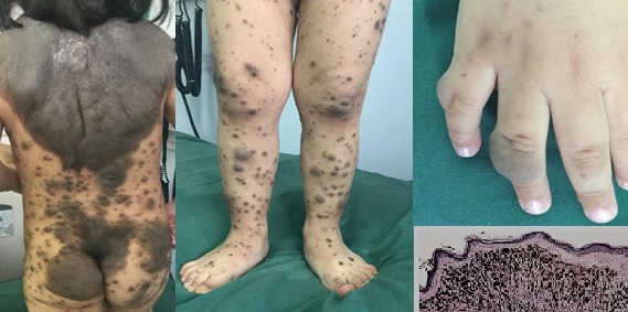
Giant congenital melanocytic nevus Case Report
Main Article Content
Abstract
Introduction: Congenital melanocytic nevi (CMN) are skin lesions that are frequently present from birth; however, the presence of a giant CMN greater > 20 cm is infrequent and forms the basis for this case report.
Case: A boy of two years and five months, who presented congenital nevi of different diameters scattered throughout the skin area, the largest being a dark-colored embossed one in the posterior thorax area along the mid-dorsal line. The nevus rose from the skin and started from the occiput and extended through the midline until it reached the sacral region and buttocks. It covered the shoulders in an inverse triangular shape with diameters of 27 by 25 cm. It was accompanied by numerous satellite nevi ranging from 3 mm to 15 cm. The presence of two neurofibromas on the fingers was noted.
Evolution: A consultation with pediatric neurology concluded in a neurological examination without alteration, the study of brain nuclear magnetic resonance and of the spinal canal, were normal, as well as the complementary tests of hematic biometry, blood chemistry, liver and thyroid profiles, and abdominal echo. The skin biopsy reported a histological pattern of melanocytic nevus. Due to the extent of the injury, observation was decided. The pruritus was treated symptomatically.
Conclusion: CMN Syndrome is associated with multiple classic phenotypic findings among which are pigmentation patterns that occupy Blaschko's lines, neurofibromas, and multiple satellite melanomas. Diagnosis is based on clinical features, and its treatment requires surgical procedures that take into account the extent of the lesion. Comprehensive management in an interdisciplinary manner is essential for treatment of CMN
Downloads
Article Details

This work is licensed under a Creative Commons Attribution-NonCommercial 4.0 International License.


